
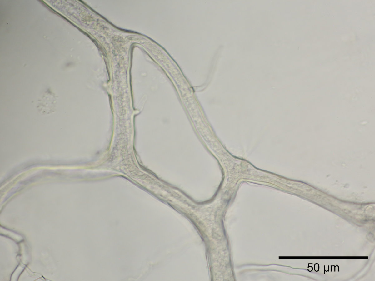
To this day, the mechanisms responsible for bidirectional streaming within hyphae remain unexplained. This page presents a series of videos that allow the phenomenon to be observed directly and hypotheses to be formed.
At high magnification, the microscope’s limited depth of field sometimes allows only a single slice of the hypha to be observed at a time. The streaming visible at the surface may therefore differ from that occurring deeper within. Each video should be interpreted with this optical constraint in mind, and full-screen viewing is recommended to fully appreciate the details.
Menu
- Introduction to Bidirectional Streaming
- Effect of Hydric Changes in the Environment
- The Plant Influences the Streaming
- Organelles Appear Able to Swim on Their Own
- Absence of a Dividing Membrane
- Bidirectional Streaming in a Loop
- Streaming in the Apex
- There Sometimes Appears to Be Pressure Within the Hypha
- Electron Microscope Photographs
- Conclusions and Hypotheses of the Author
- Additional Information from Other Researchers
- Dr. David Naylor’s Video
Introduction to Bidirectional Streaming
Hyphae of arbuscular mycorrhizal fungi appear to be traversed by a cytoplasmic current carrying organelles that circulate simultaneously forward and backward within a single hypha. In every species observed so far, this streaming is markedly faster than the very slow movement seen in septate fungi (Basidiomycetes, Ascomycetes, etc.).
The phenomenon is as follows: cytoplasm and organelles appear to circulate simultaneously in both directions — forward and backward — within a single coenocytic hypha, without any membrane or separating structure ever having been identified, even under electron microscopy. A dominant flow in one direction is often observed, with a secondary flow in the opposite direction: sometimes arranged left-right, sometimes center-periphery. The apparent speed of organelles moving “forward” often seems to differ from that of organelles moving “backward.” It should be noted that the cytoplasm is normally transparent, so what can be seen moving are the organelles — the actual movement of the cytoplasmic fluid itself remains invisible, which of course adds to the mystery.
Most of the videos below feature Rhizophagus irregularis, always in symbiosis with a normal host plant.
The introductory video begins with the stereo microscope on a hyphal segment, then gradually zooms in under the microscope to make the bidirectional streaming clearly visible. The video runs two minutes and loads (slowly) from an external server — please allow some time at startup.
A second video illustrates two simultaneous types of streaming: left/right in the hypha on the left, and center/periphery in the upward-oriented hypha. It also shows unidentified “blue balls,” occasionally present in mature Rhizophagus irregularis hyphae.
*This video also appears in the “Filming Cytoplasmic Streaming in Hyphae” section.
Dr. David Naylor (Toronto Metropolitan University), a fluid mechanics specialist, produced a very interesting YouTube video based on this footage; a link to it appears at the bottom of the page.
Effect of Hydric Change
This observation involves a Diversispora varaderana hypha in active symbiosis with Plantago lanceolata, in a Petri dish that had not been irrigated for 15 days. Both the plant and the fungus were therefore moderately dehydrated. Placing the dish on the inverted microscope makes it possible to observe actively streaming hyphae on the dish’s dry floor, between two polymer grains. A syringe allows the dish to be discreetly irrigated through its openings without disturbing it.
The following observations can be drawn from this:
- The speed of streaming can be strongly affected by hydric changes in the environment.
- The direction of streaming can reverse within a few seconds following a hydric change.
- The cytoplasmic flow can become strongly unbalanced between the two directions.
The Plant Influences the Streaming
This video strikingly demonstrates that the two symbionts influence each other in real time. It features Rhizophagus irregularis in symbiosis with Plantago lanceolata. The plant’s stem is cut at the crown at the 28-second mark of the video, while the Petri dish rests on the inverted microscope. Within a few seconds, the direction of streaming reverses. The original video runs nearly 6 minutes to document the stability of streaming before and after the cut; only a short segment is shown here, sped up 4×. At the start, the main current flows to the left and downward; after the cut, it reverses toward the right and upward.
It is remarkable that an action performed on the plant is reflected in the fungus within mere seconds.
Organelles Appear Able to Swim on Their Own
This first video shows a constant flow of organelles entering and exiting two closed, spherical spores. It is difficult to attribute this movement to a simple cytoplasmic flow exiting closed structures. These organelles appear to move autonomously, with some circulating against the current. By what mechanism do they orient themselves? The question remains open. This is Rhizophagus irregularis in symbiosis with Plantago lanceolata, filmed in real time (1×).
In the following video, material (organelles, and presumably cytoplasm as well) enters through the hypha, turns around, and exits through that same hypha. How can this be explained?
Absence of a Dividing Membrane
Erratic movements can be observed within the various flows inside hyphae, particularly where flows meet. If a membrane separated flows moving in opposite directions, movements should be far more clearly oriented. The following video strongly suggests that no membrane separates these flows.
This video first shows the apex of Funneliformis geosporus, followed by a close-up of a short section posterior to this apex. In both cases, no septal membrane is evident. Numerous active bacteria are also observed around the hypha, a phenomenon commonly seen in non-sterile culture media.
Bidirectional Streaming in a Loop
Streaming is not only typically bidirectional, but it can also organize itself into a loop. This type of configuration has been observed on several occasions. The following video presents a particularly well-defined example. It features Rhizophagus irregularis in symbiosis with Plantago lanceolata, in real time (1×).
Streaming in the Apex
To grow, a hyphal apex must continuously receive the components needed for cell wall synthesis (lipids, proteins), as well as a source of energy (ATP, mitochondrial activity, etc.). The following video shows active streaming at the apex of a growing Gigaspora rosea hypha. Since these cultures are grown in non-sterile conditions, bacteria and other microorganisms can be seen in the periphery — which is entirely normal in this context.
There Appears to Be Pressure Within the Hypha
Certain observations suggest that streaming follows a pressure gradient. The following video offers a particularly illustrative example, with zoom and slow-motion footage.
Electron Microscope
Here are a few photos of Rhizophagus irregularis hyphae taken under an electron microscope. No evidence of a membrane separating compartments can be seen.

©Sa Majesté le Roi du chef du Canada, représenté par le ministre de l’Agriculture et de l’Agroalimentaire
©His Majesty the King in Right of Canada, as represented by the Minister of Agriculture and Agri-Food

©Sa Majesté le Roi du chef du Canada, représenté par le ministre de l’Agriculture et de l’Agroalimentaire
©His Majesty the King in Right of Canada, as represented by the Minister of Agriculture and Agri-Food
Conclusions and Hypotheses of the Author
Taken together, these observations point to the hypothesis of two complementary mechanisms:
First mechanism — pressure on the cytoplasm: a mechanism generates pressure on the cytoplasmic fluid, forcing it to flow in one direction or the other. This mechanism appears sensitive to the osmotic pressure of the surrounding medium as well as to signals originating from the host plant.
Second mechanism — organelle motility: organelles may be capable of moving autonomously, at a moderate speed, either with or against the cytoplasmic current.
Bidirectional streaming could thus be explained as follows: some organelles are carried along by the current, while others swim against it. During episodes of strong cytoplasmic flow, all organelles are swept along in the same direction.
Naturally, these are not “robust” conclusions but rather hypotheses grounded in recurring observations. They represent the author’s best interpretation to date, pending further work by other researchers to confirm or, alternatively, disprove them.
Additional Information from Other Researchers
Scientific Publications on This Topic:
A recent publication (2026) offers a rigorous scientific perspective on the subject: Cytoplasmic flow dynamics in arbuscular mycorrhizal fungi are intrinsic and independent of plant hosts.
This article (2025): Fluid mechanics within mycorrhizal networks: exploring concepts, traits, and methodologies est aussi fort intéressant.
Dr. David Naylor’s Video
Dr. David Naylor, a fluid mechanics specialist, is warmly thanked for this wonderful contribution.