
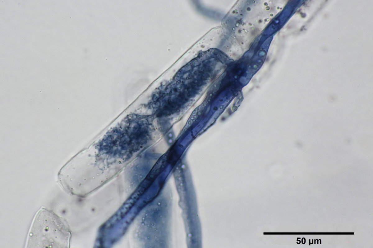
Arbuscule of Funneliformis geosporus, stained using the Ink and Vinegar technique.
This fungus proves fairly easy to grow and multiply. It produces an abundance of hyphae in the polymer as well as numerous spores, both in the polymer and in the vermiculite. A new generation can be started without difficulty from polymer grains containing spores, and the culture establishes itself quickly.
Three types of spores are often distinguished in the polymer: 1) large yellow spores, typical of the species; 2) large white spores, whose nature remains uncertain — they could be immature spores or a different structure; and 3) small whitish spores, often partially empty, whose origin also remains to be determined. Regular photographic monitoring would likely help clarify this question; it represents a promising avenue for further observation.
Menu
- 33-Day Survival in Refrigeration
- Comparison: Commercial 30-10-10 Fertilizer vs. M2P
- Microscopy
- Aerobic Fungus
- Notes on an Eight-Month-Old Culture
33-Day Survival in Refrigeration
Three Plantago lanceolata plants colonized by F. geosporus survived a thirty-three (33)-day stay in the dark in a refrigerator at 4 °C.
An extended absence from the lab, with no one to look after the cultures, gave rise to an improvised experiment: three (3) dishes of P. lanceolata well colonized by F. geosporus were placed in a well-sealed Ziploc freezer bag, then stored in the refrigerator. The plants were expected to die, with the intention of reusing the colonized roots upon return to restart the cultures. These three plants had been in culture for three (3) months; root density in the dish was therefore high, and the plants were in excellent condition. The fungus was likewise abundant in the polymer.
Against all expectations, the three plants survived this 33-day stay and resumed growth as soon as they were returned to regular grow lighting (LED). The old polymer was then removed and replaced with fresh, uncolonized polymer to check whether the fungus was still active. An observation made 46 days after this replacement confirms that all three plants are healthy, with normal foliage. The fungus intensely recolonized the entire polymer and produced an abundance of new spores: both the plant and the fungus are clearly thriving.
This result leaves no doubt: the roots or spores are enough to restart a new generation. Here are two images of the new polymer:


Comparison: Commercial 30-10-10 Fertilizer vs. M2P
A comparative performance trial between a commercial 30-10-10 fertilizer and the M2P solution was carried out with F. geosporus and R. irregularis. Details are described on the miscellaneous experiments page, accessible here. Here is the conclusion:
Plant growth was very rapid, and the dishes irrigated with 30-10-10 dried out (too) quickly, which appears to have harmed F. geosporus, which seems less “fast” than R. irregularis at colonizing roots. Once a root is colonized, both fungi grow rapidly and tolerate drought well. Several F. geosporus dishes showed a marked delay in colonization, likely due to drought; once that issue was resolved, hyphal and spore production returned to normal.
The F. geosporus dishes had been inoculated with colonized polymer grains, and this inoculum proved effective.
Microscopy
The small white spores are difficult to classify. Many small white spores and many large orange spores are observed, but few intermediate ones. The small white spores often appear half-empty or partially transparent — a promising subject for further study. The hyphae, meanwhile, display the typical bidirectional streaming, which is easy to observe.

Staining with the Ink and Vinegar method works well with this fungus: it allows hyphae, vesicles, and arbuscules to be easily distinguished.


Aerobic Fungus
F. geosporus appears to show partially aerobic behavior. The following photo illustrates an eight (8)-month-old dish: the fungus produced numerous spores in aerated conditions there, on and within the vermiculite, as well as on the surface and inside the polymer grains. The vermiculite surface inside the dish forms a humid micro-environment, since the lid remains in place at all times. Note that some spores, not shown here, occur directly on the roots.

Long-Term Culture: Eight Months
Growing Funneliformis geosporus with the host plant Plantago lanceolata can easily extend over eight months, and probably well beyond. Several dishes of P. lanceolata were inoculated with colonized polymer grains in September 2025 to compare different fertilizers. At the end of the experiment, a few dishes irrigated with M2P were simply kept until May 2026, or roughly eight (8) months. The plants are still doing very well, and in March 2026, a few colonized grains were used to successfully start new cultures.
At this stage, after eight months of culture, observation of the three remaining dishes reveals the following:
Spores are found throughout the dishes (not illustrated): spores are observed on the polymer side, within the grains themselves and on the plastic floor of the dish. On the vermiculite side, they are found on the surface as well as around the roots, when the dish is turned over. This suggests the vermiculite likely also contains large numbers of spores, although extraction was not performed: colonized polymer grains are generally sufficient to restart cultures.
Hyphal density can become very intense, almost “cottony” given how many hyphae occupy certain portions of the dish, especially near the dividing membrane. Hyphal density is not uniform; it is high near the dividing membrane and decreases with distance from it.

Many hyphae are empty. The large majority of them are empty and often sealed off by a small internal wall. Considerable searching is needed to find a hypha that is still active.

Some hyphae have become very thick. The oldest hyphae are probably the thickest: no hypha this thickened has ever been observed in a young dish, suggesting they thicken over time. Here, the thick hypha is empty, but thick hyphae with very active streaming have also been observed.

No vesicles are observable. Direct observation under the microscope or stereo microscope does not reveal any vesicles or arbuscules in living roots, unlike R. irregularis, where vesicles are often quite apparent.
Contaminant density has very little effect on observations and does not appear to harm the cultures either. At high magnification, motile bacteria can easily be observed, along with protists that likely feed on them. The exact origin of these contaminants is unknown, but they are present more or less everywhere in the air and on materials.
A light presence of microscopic algae is also often observed on the vermiculite, wherever it is exposed to light; these algae, however, do not reach the polymer.
The identity of contaminants is usually unknown, and there are potentially thousands of possible contaminant species. Some contaminants could certainly harm the cultures, but in general, the cultures appear to withstand their presence well.