
Menu
Ink and vinegar
The technique developed by Vierheilig — Ink and Vinegar, a Simple Staining Technique for Arbuscular-Mycorrhizal Fungi — has the advantage of not requiring any carcinogenic chemical products. The version used here is a simplified adaptation of this method:
- Prepare the 10% (wt/vol) KOH solution as described in the method. To keep it simple, if you dissolve 10 g of KOH in 100 ml of water, this solution will work very well. Always add the dry KOH gradually to the liquid, never the other way around.
- Prepare the black Sheaffer ink no. 94231: 5 ml of ink in 95 ml of 5% vinegar. Regular grocery-store 5% vinegar works well.
- Place the roots for 3 minutes in the KOH solution preheated to between 90°C and 100°C, then rinse for a few minutes in a beaker with tap water
- Place the roots for 3 minutes in the ink-vinegar solution preheated to between 90°C and 100°C, then rinse for a few minutes in a beaker with tap water.
- After that, it is ready for observation. Sometimes it is preferable to let it destain overnight, but in general, excellent observations can be made immediately after rinsing.
I detail at this link a tip for staining small volumes of roots.
Here is some additional information about the method:
- Treatment times in the KOH and vinegar solutions vary depending on root thickness, their cellulose content, plant species, etc. Trials must be done on a case-by-case basis. The times described here work well for Plantago lanceolata. Other plants might require more time.
- To save time, I use the microwave to raise the temperature of the solutions to 90°C, and then place the beaker on the hot plate in order to better control the boiling and stir the solution.
- For an unexplained reason, this technique does not seem to stain the fungus Diversispora varaderana.
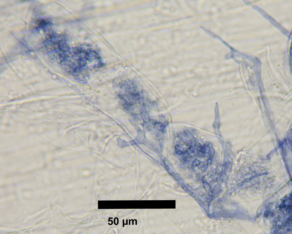
Here are some photos of the results


Regarding the superabsorbent polymer in the KOH and vinegar:
To date I have often put polymer grains in the KOH and then in the boiling vinegar without observing any degradation or breakdown of the polymer. APPARENTLY, the polymer seems inert and does not react chemically, although it shrinks a little. The ink penetrates the grains and comes back out when they are placed in water. Precise chemistry work would be needed to guarantee the polymer’s complete safety under these conditions, but I am not aware that this has been done. There is therefore no absolute certainty about the reactions or absence of reactions of the polymer in this process. Proceed carefully according to good laboratory practices. If in doubt, do not hesitate to work under a chemical fume hood.
Methylene blue on live roots and hyphae
This is a simple “let’s see” test. In a petri dish containing P. lanceolata and R. irregularis, with the polymer in fine suspension (see here how to make it), I placed a drop of methylene blue (1% aqueous solution) to see the staining effect. The dye quickly affects all the material it touches and then diffuses slowly into the polymer. The observed effects are:
- 1) The roots are stained lightly pale blue
- 2) Some root cells (dead?) are strongly stained in a regular “pattern”.
- 3) The dye appears to be transported internally by the roots, since stained roots can be seen far from the diffusion zone in the polymer
- 3) Some spores, but in a small proportion, are stained, likely because they are dead, as I no longer detect any movement in the suspensor hyphae. The stained spores are located where the drop landed.
- 4) The extraradical hyphae are not stained
- 5) Neither the intraradical hyphae nor the vesicles are stained
In conclusion, methylene blue does not reveal much about the fungus. However, the pattern of dead cells on the roots is intriguing. Does the fungus play a role in the death of these cells?


extraradical hyphae are not stained and no intraradical hyphae are visible..
Sheaffer ink on live roots and hyphae
This is a simple “let’s see” test. In a petri dish containing P. lancolata and R. irregularis, with the polymer in fine suspension (see here how to make it), I placed a drop of pure black Sheaffer 94231 ink (no dilution) to see the staining effect. The dye quickly affects all the material it touches and then diffuses slowly, over several days, into the polymer.
The staining reaches all the spores, but did not stain the suspensor hyphae. . It seems that the “runner” hyphae carried the dye inside the most distant spores, and that the dye accumulates there enough to become noticeable.
The observed effects are:
- 1) After the drop is placed, a black spot forms within a few seconds and spreads little
- 2) The ink diffuses slowly, over several days
- 3) The spores are stained black after a few days, even those that appear far from the still-tinted polymer
- 4) The suspensor hyphae and the small hyphae do not seem to get stained, or only very slightly.
- 5) Some hyphae, notably the “runner” hyphae, seem to become darkly stained. These hyphae show active circulation despite the staining.
- 6) No intraradical structure appears to be stained or revealed
Conclusion: Ink used in pure form does not seem to provide much new information. However, the staining of the “runner” hyphae suggests that hyphae transport the dye to carry it inside the spores.


